Explaining the changing shapes of enzymes
Distinguished University Professor lecture
To learn what a protein does inside a cell, you need to know how it looks. Every molecular twist and turn, every chemical loop and ring has a meaning all its own. Even minor variations in protein structure can produce major changes in function. Just to make things even more complicated, many proteins change their shapes when they bind to or interact with other molecules.
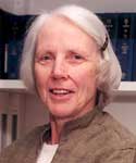
(Photo courtesy Martha Ludwig)
Martha Ludwig, the J. Lawrence Oncley Distinguished University Professor of Biological Chemistry, has devoted her long and distinguished career to probing the secrets of protein structure using a technique called X-ray crystallography.
She joined the faculty in 1967, and now is a professor of biological chemistry in the Medical School and a research scientist in the Biophysics Research Division.
“The visual part of my work is personally satisfying to me,” Ludwig said in an article distributed by the National Academy of Sciences soon after she was elected a member of the organization in April 2003. “I’m always delighted to look at a new three-dimensional protein structure. Many of our basic hypotheses of how proteins work have come directly from observing structures.”
Ludwig will deliver her Distinguished University Professor lecture, “Conformational Changes in Catalysis: Enzymes as Molecular Acrobats,” at 4 p.m. Feb. 1 in the Rackham Amphitheatre, followed by a reception in Assembly Hall.
Enzymes are proteins that serve as catalysts during biochemical interactions. Without enzymes, most of the basic functions vital to life would not be possible. Ludwig will focus on three specific research studies in her lecture, because they provide examples of the connection between an enzyme’s structural flexibility and its function.
In a group of proteins called flavodoxins, for example, the position of the peptide backbone in the enzyme has been shown to control electron transfer between the enzyme and its cofactor. In another enzyme called p-hydroxybenzoate hydroxylase (PHBH) the flavin cofactor moves back and forth between alternate positions to support catalysis of different reactions. Dramatic rearrangements in the structure of an enzyme called B12-dependent methionine synthase are used to move vitamin B12 to several different sites involved in the synthesis of methionine.
In her lecture, Ludwig also plans to credit several U-M scientists who were her mentors and had a profound influence on her research and career—especially Oncley, a former professor of chemistry and biological chemistry and first director of the Biophysics Research Division, and Vincent Massey, a former professor of biological chemistry in the Medical School.
“My research has benefited enormously from the rich intellectual environment at this University, and has been enabled by collaborations and interactions with many colleagues and students,” she says.